-
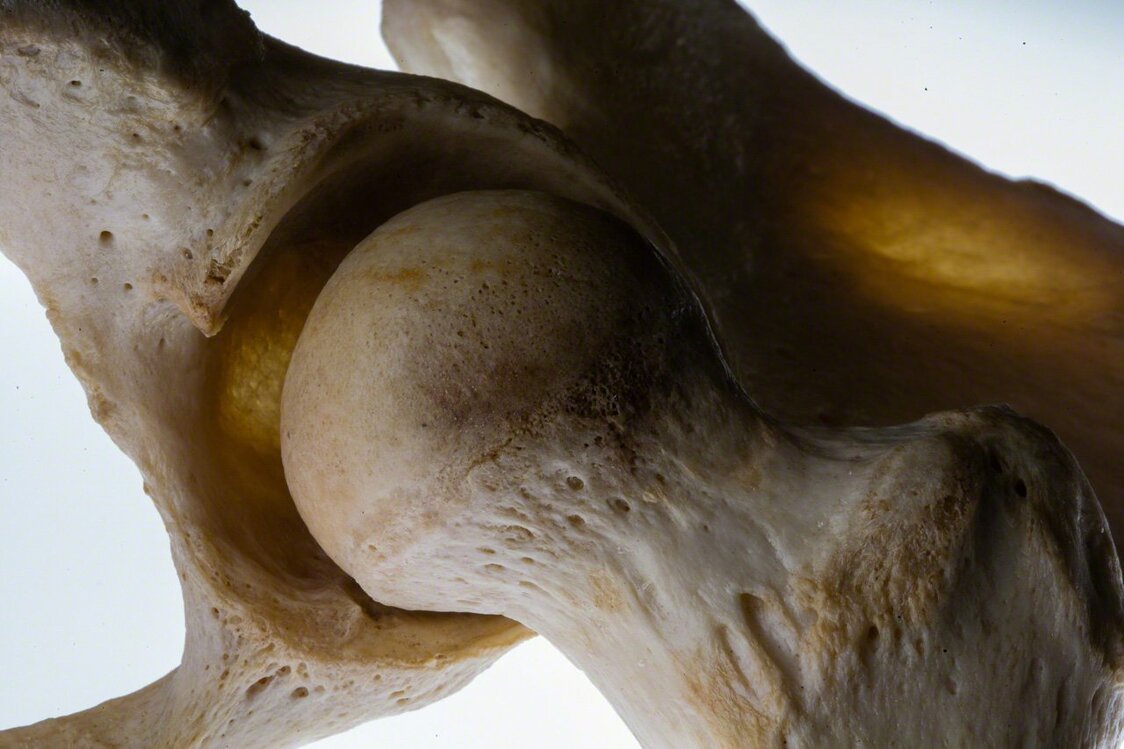
The head of the femur, or thigh bone, fits into and swivels freely inside the cup-shaped acetabulum. (© Ōnishi Naruaki)
-
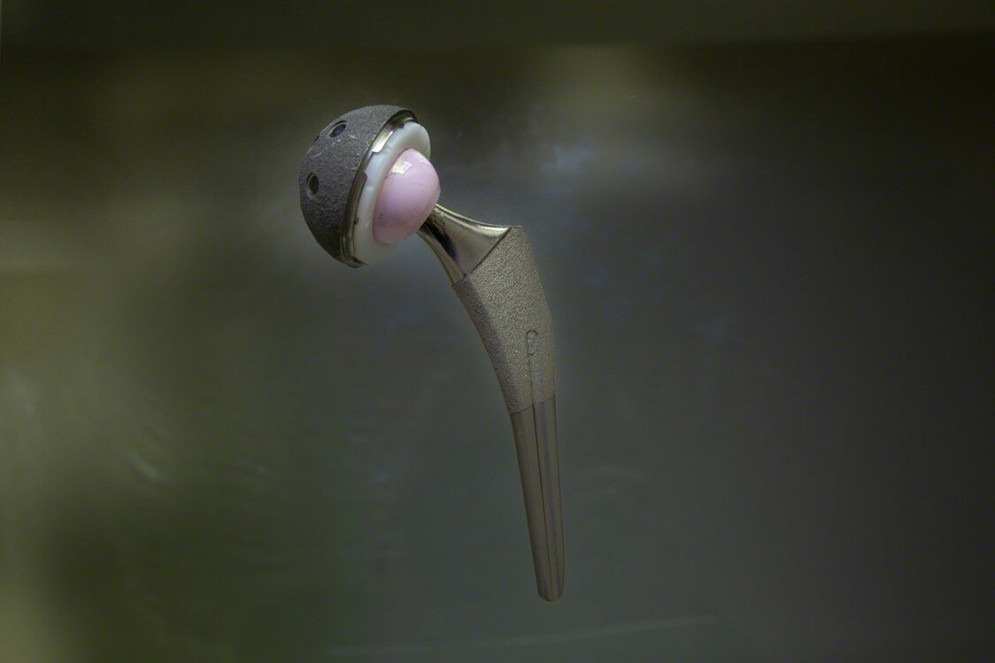
A hip implant system manufactured by the US firm Stryker. The implant weighs about 100 grams more than the bone and cartilage it replaces. (© Ōnishi Naruaki)
-
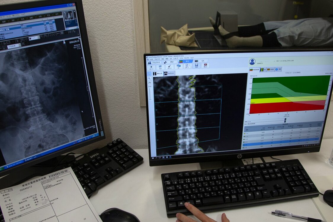
A bone density scan is part of the battery of preoperative tests conducted before hip replacement surgery. (© Ōnishi Naruaki)
-
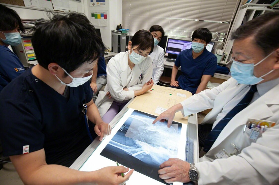
The medical team gathers to draw up a personalized surgical plan on the basis of the patient’s test and imaging results. (© Ōnishi Naruaki)
-
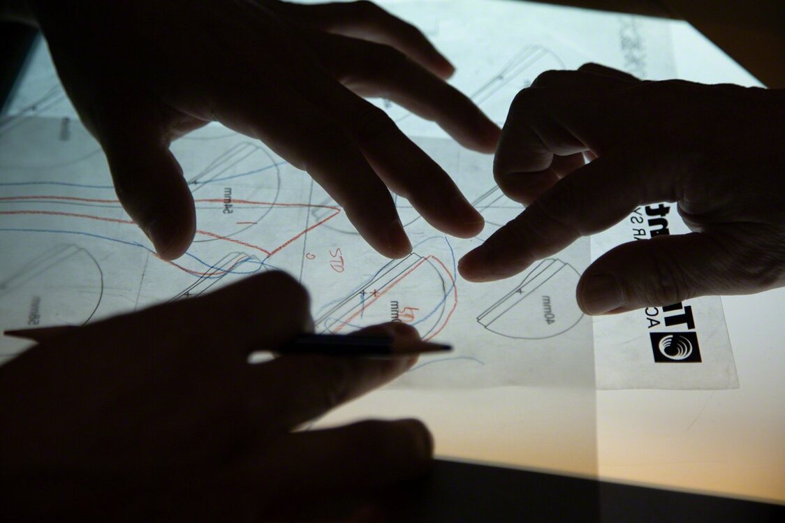
Using a protractor and straight-edge, the surgeon draws a precise diagram of the implant in relation to the patient’s anatomy. The exercise helps imprint the surgical plan on the surgeon’s physical memory. (© Ōnishi Naruaki)
-
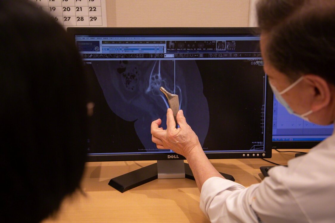
“This is how we’ll insert the hip implant,” explains Dr. Matsubara. He spares no time or effort ensuring that each patient understands the procedure and is comfortable with it. (© Ōnishi Naruaki)
-
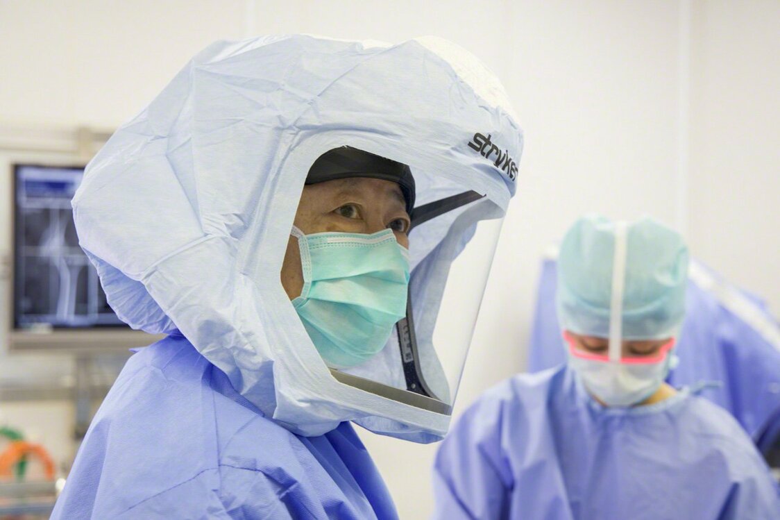
An extra layer of personal protective equipment, including a surgical helmet, is used to guard against infection, a major concern when implants are used. Matsubara jokes that he became an orthopedic surgeon so that he could dress up in a space suit. (© Ōnishi Naruaki)
-
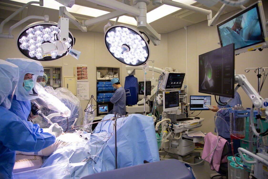
The Mako robotic-arm assisted surgery system springs into action. Dr. Matsubara guides the robotic-arm as the attachment spins, removing bone tissue. The monitor at right shows the section to be removed in green. A clean removal leaves only white. Any excess removal appears in red. If the cutting proceeds more than 2 mm beyond the predetermined boundary, the arm automatically stops. (© Ōnishi Naruaki)
-
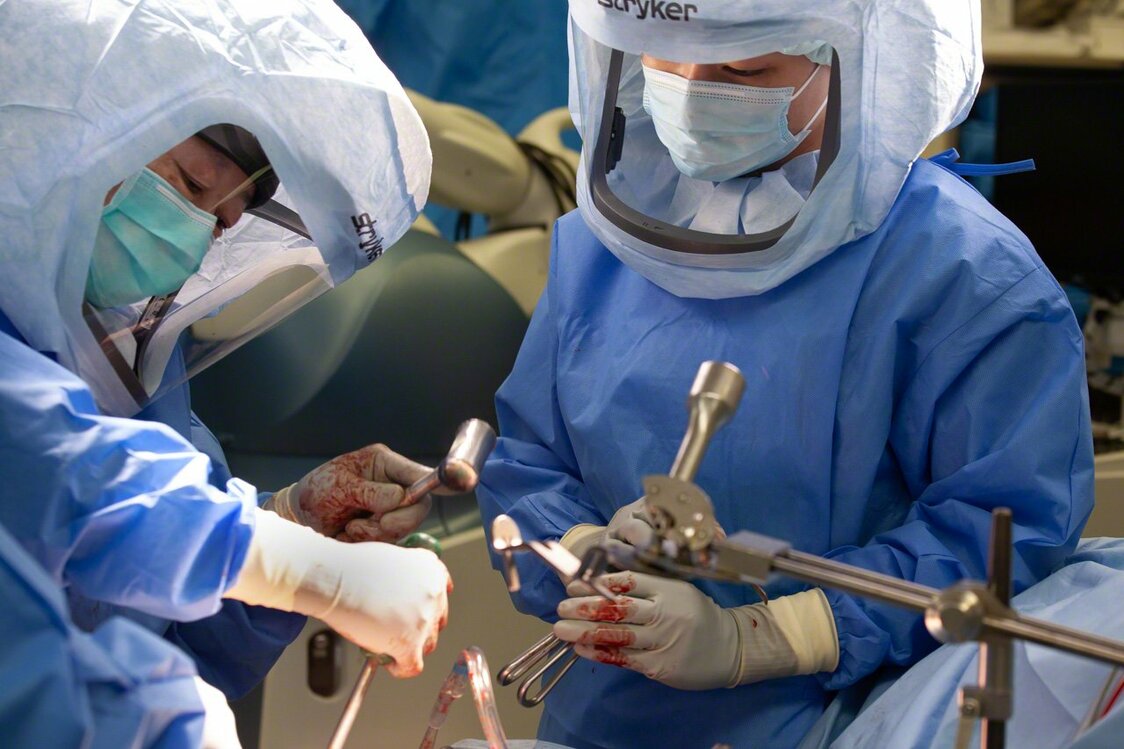
Dr. Matsubara uses a mallet and introducer to seat the artificial acetabular cup in the acetabulum. The surgeon can hear and feel the cup pop into place when the angle and depth of insertion are just right—a more reliable indicator than any electronic gauge. (© Ōnishi Naruaki)
-
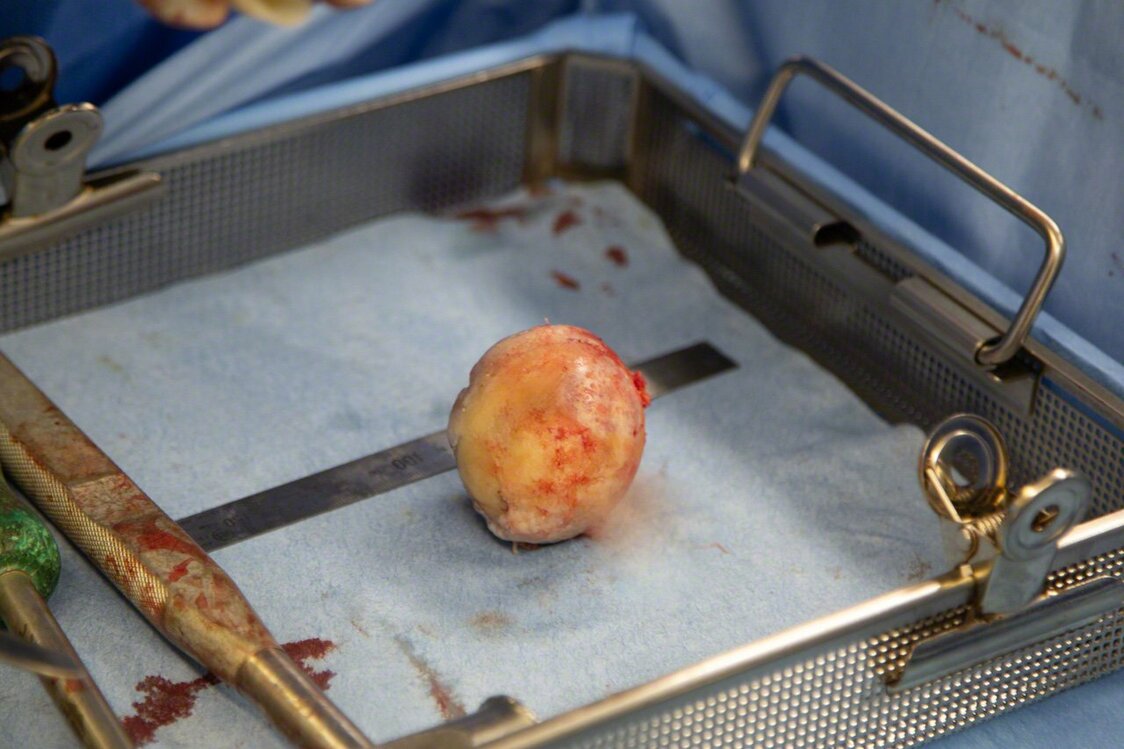
My wife’s surgically removed femoral head. The cartilage, discolored from disease, had worn down to the bone in spots. (© Ōnishi Naruaki)
-

The surgical team examines a set of X-rays to confirm correct installation of the implant, the final step in a procedure lasting just 1 hour and 17 minutes. (© Ōnishi Naruaki)
-

Rehabilitation begins the day after the operation. “What patients need following this surgery is not rest but movement,” says the physical therapist, shown guiding my wife through range-of-motion exercises. (© Ōnishi Naruaki)
-
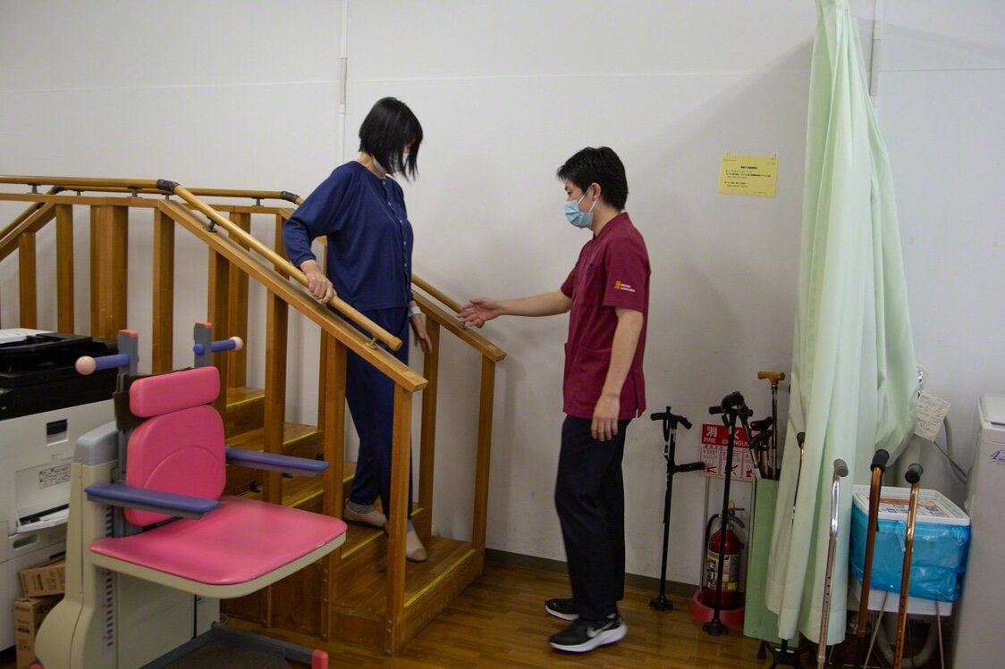
Before discharge, the patient receives occupational therapy tailored to her individual living environment, hobbies, and other demands. Thanks to this support, my wife was able to come home eight days after undergoing surgery. Full recovery will take time, but her face lights up as she talks about the places she wants to go and the things she wants to do in the coming years. (© Ōnishi Naruaki)
-
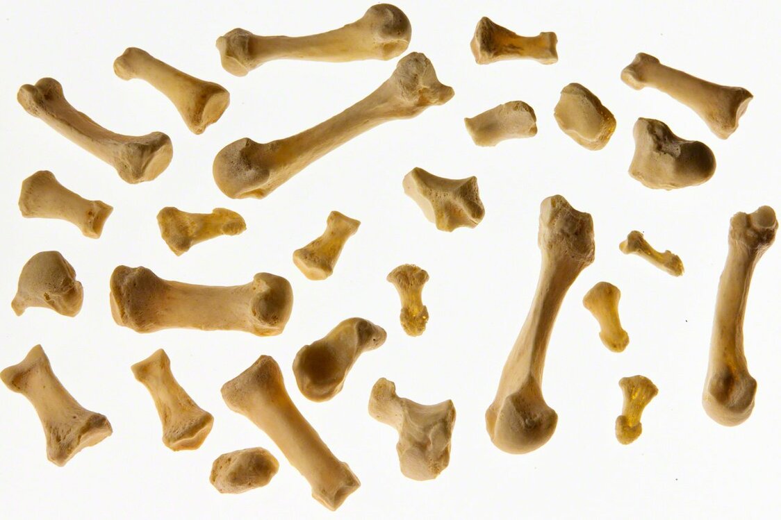
There are 206 bones in the adult human skeleton, and every one is unique.(© Ōnishi Naruaki)


 Read in other languages