-
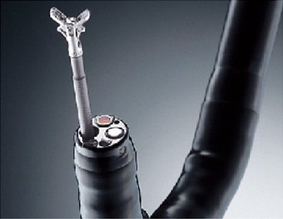
A lens, a CCD image sensor, and two lights lie at the end of the endoscope. Two lights are used to prevent tools protruding from the end of the endoscope from casting a shadow. Tools are inserted into a channel that can also be used to suck fluids out of the patient.
-
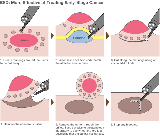
ESD is the latest method of surgery performed with endoscopes. Previously, EMR (endoscopic mucosal resection) procedures used a lasso-shaped tool to cut away tumors, but could only deal with growths below a certain size. By using a small scalpel, doctors can use ESD to excise tumors larger than 2 centimeters. In Japan, around one in four stomach cancer patients are already receiving this treatment. Source: “Gan o kirazu ni naosu naishikyō chiryō ESD ga wakaru hon” (Understanding ESDs: A Procedure for Treating Cancer Without Major Surgery) (Kōdansha, 2011).
-
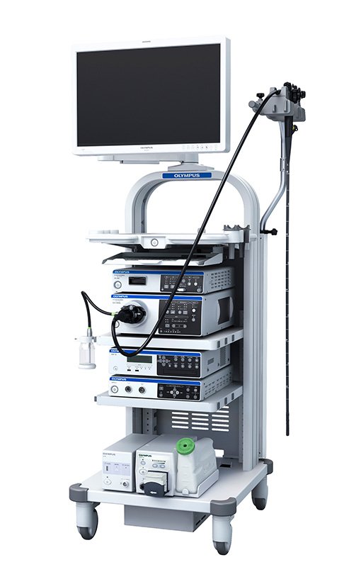
The video scope system primarily consists of a unit that sends light to the end of the endoscope, a video processor that takes the images from the CCD and displays them on the monitor, and other components. Xenon lamps, a particularly powerful light source, are used to get a clear picture from the dark spaces inside the body.
-
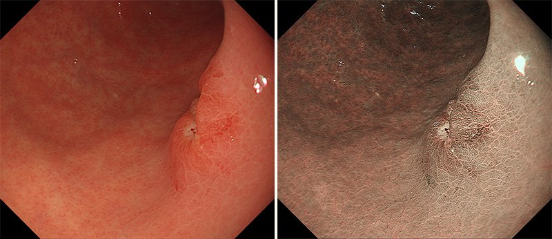
With normal light, the blood vessels are drowned in the pink color of the internal organs, making them more difficult to discern (left). Blue light (415 nanometer wavelength) and green light (540 nanometers) make the blood vessels much easier to see. (Photos: Kawamura Takuji, Department of Gastroenterology, Kyoto Second Red Cross Hospital.)


 Read in other languages